Common Dental Problems
 What is under all of that tartar?
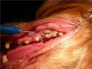
Brittney presented for a routine dental cleaning. When all of the tartar was removed from the upper left fourth premolar, a slab fracture of the crown and root was found. This fracture had allowed bacteria to infect the tooth root area, undoubtedly causing discomfort. Dental x-rays found there to be an abscess of the root associated with this tooth. Despite this, she never showed her owners any signs of pain or discomfort and continued to eat normally (although probably using the other side of her mouth)! The tooth was extracted as part of the dental cleaning and healed without problem. No change in diet was needed except for soft food in the first few days after the procedure.
 How Old Is Too Old For A Dental Cleaning?
Although this is not a procedure to be taken lightly, it can routinely be done safely in the very old if (1) bloodwork shows no significant underlying health problems, (2) there are no underlying heart problems, and (3) anesthesia is performed in a thoughful and careful manner with aggressive anesthetic monitoring and supportive care.
Molly is a geriatric Lhasa Apso that had terrible dental disease and many rotten teeth. Her teeth had never been cleaned due to the owners concerns regarding anesthesia. Given the severity of dental disease present during examination, the owner elected to have her teeth cleaned and the many rotten teeth pulled. Two weeks after the procedure, Molly was rechecked and the owner indicated that she was acting like a puppy for the first time in a long time.
 Feline Odontoclastic Resorptive Lesions
Cats have a unique dental disease called feline odontoclastic resorptive lesions (FORL). This is an inflammatory disease process whereby the tooth actually resorbs, often at or below the gum line, and eventually breaks. FORL’s are painful and sometimes signs are noted including drooling, bleeding, and difficulty chewing. Although they are in pain, many cats do not exhibit these clinical symptoms and the lesions are found on routine examination.
 An oral examination of the cat’s mouth sometimes reveals a cherry-red inflammation of the gums surrounding the affected tooth. FORL’s can also be demonstrated by gently rubbing the suspected lesion with a cotton swab. If the lesion is present, pain and jaw spasms occur when the area is touched.
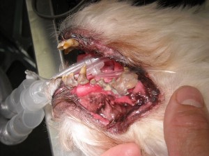
Princess presented for a dental cleaning. Upon examination, the right side of her jaw (pictured on the left) had a large amount of proliferative gum tissue over the area of the second molar. Dental x-rays showed that the crown had broken off and roots were still present, likely causing her pain and discomfort. This was a late stage FORL.
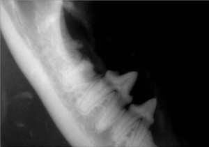
Upon visual examination of the left side of the jaw, the second molar was noted to be partially broken apart by a FORL. There was nothing that could be done to save the tooth. The X-ray (pictured on the right) shows one root and part of the crown to the second molar present while the second root had already been broken down by the body. The dental x-rays were helpful in identifying what part of the tooth needed to be extracted, limiting trauma to the mouth and discomfort to the patient.
 Dental X-rays Tell A Greater Story
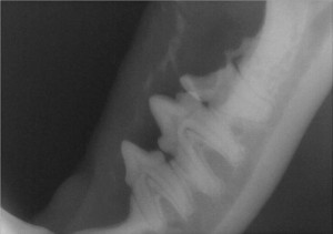
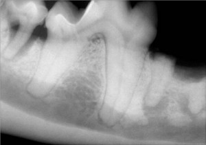
The center tooth in this x-ray is how a normal tooth and its roots should appear. The bone around a tooth root should always appear white and smooth. Black areas are an indication that bone is lost, which occurs with infection and abscessation.
The tooth in the far left has vertical bone loss around the tooth root as demonstrated by the darker area around the tooth root. This bone loss occurs when bacteria associated with tartar infect below the gum line. This is periodontal disease.
The tooth on the right has lost the crown (the top portion from an earlier fracture that went unnoticed. With time, bacteria traveled through opening in the center of the tooth and infected the apex (tip). This has resulted in the dark pocket on the x-ray around the apex. This is a tooth root abscess.
It is interesting to note that this dog did not show any signs to the owner even though, at times, she was clearly experiencing pain.